Asheq, PhD student, joins lab

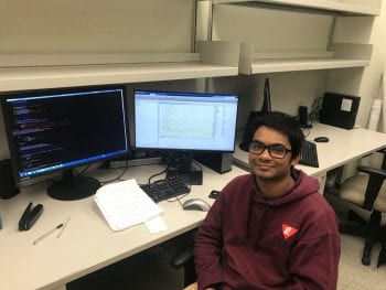
Excited to share that Md Ashequr Rahman, PhD student from the Image Science PhD program, has joined the Computational Medical Imaging Lab. Asheq obtained his Bachelors in Electrical and Electronic Engineering from Bangladesh University of Engineering and Technology, Bangladesh. Welcome Asheq!

Dr. Jha delivers keynote at Siteman Oncologic Imaging program

Dr. Abhinav Jha presented the keynote at the Siteman Cancer Center Oncologic Imaging Research Program Student Poster Session and Special lecture. His talk was titled  “Computational Medical Imaging for Precision Medicine” and discussed various computational-imaging, that included machine-learning and model-based methods, to improve precision medicine. Further, excited to share that Priyanshu Jain, a student member […]

NIBIB Trailblazer award on SPECT reconstruction

Our lab has been awarded the NIBIB Trailblazer award for a project on developing new methods for transmission-less attenuation compensation in SPECT imaging! In this project, we propose to design and validate novel methods for SPECT reconstruction that will not require an additional transmission scan (such as the CT scan) for attenuation compensation. For more […]

NVIDIA GPU grant

Our lab has been awarded the NVIDIA GPU grant! NVIDIA will provide us state-of-the-art Quadro P6000 card. Armed with 3,840 CUDA parallel processing cores, a peak single-precision performance of 12 TFLOPS, and a 24 GB GPU memory, this card will provide an excellent high-performance computing platform for several computational medical imaging solutions that we are […]

New paper on reconstruction method for FMT brain imaging

Our paper on developing a new reconstruction method for fluorescence molecular tomography in the context of transcranial imaging has been published in Biomedical Optics Express. The paper demonstrates that accurately modeling noise in FMT while accounting for sparsity leads to improved reconstruction performance!

Abstract on deep learning for PET image analysis accepted @ SNMMI

Our abstract titled “A deep-learning-based fully automated segmentation approach to delineate tumors in FDG-PET images of patients with lung cancer” was accepted for an oral presentation at the 2018 SNMMI Annual Meeting. In this work, we developed and demonstrated the efficacy of a deep learning-based method for tumor segmentation in PET images. Since these images are […]

Joining WashU as Assistant Professor of BME/Radiology

Dr. Abhinav Jha will be joining Washington University in St. Louis as an Assistant Professor of Biomedical Engineering in the School of Engineering and Applied Sciences and of the Mallinckrodt Institute of Radiology at the School of Medicine starting April 2018. His recruitment is part of the Imaging Sciences Initiative at WashU.

Three articles on developing observers for myocardial SPECT

Optimizing myocardial perfusion SPECT systems and methods for perfusion defect detection is highly significant. While mathematical observers exist for this purpose, they do not account for the variability in cardiac defect characteristics. In a series of articles, we investigate the causes for this issue and propose a novel mathematical observer that addresses this issue. X. […]

Two new journal articles on clinical quantitative PET

Metrics derived from quantitative PET, in particular intra-tumor heterogeneity, have sparked considerable interest as biomarkers for prognosis and predicting therapy response. In collaboration with radiologists at the Division of Nuclear Medicine, we have been investigating the role of intra-tumor heterogeneity in patients with different cancers. We recently published a couple of articles based on our research, […]

Three talks at SNMMI Annual Meeting 2017

We have had three talks accepted for presentation at the Society of Nuclear Medicine and Molecular Imaging (SNMMI) Annual Meeting 2017. A. K. Jha, C. Marcus, R. Wray. R. Subramaniam, and E. Frey, “A no-gold-standard framework to evaluate FDG-PET tumor-segmentation methods on the task of measuring prognostic biomarkers for lung-cancer treatment”, Therapy Center Young Investigator Award, […]

New article on ADC estimation in diffusion MRI

Our paper on developing a statistical technique to estimate a single ADC value from a lesion using diffusion MRI was recently published in the journal Magnetic Resonance in Medicine. Especially exciting that the proposed method was rigorous, computationally fast, easy-to-implement, convenient-to-use, and more accurate than state-of-the-art methods in clinical settings! This work was in collaboration with […]